Introduction: Confronting the Challenge of Calcified Plaque
In the world of interventional cardiology, the mission is clear: restore blood flow to the heart muscle by opening blocked coronary arteries. For decades, balloon angioplasty and stenting have been the gold standard, saving countless lives. But what happens when the enemy—the plaque clogging the artery—isn’t just soft and fatty, but hard as rock?
This is the challenge of severely calcified coronary artery disease. Imagine trying to push a scaffold (a stent) through a concrete pipe. A standard balloon might not even inflate properly, and if it does, the stent may not deploy symmetrically or fully expand. This increases the risk of complications both during and after the procedure, including stent failure and re-narrowing of the artery.
To tackle this formidable adversary, cardiologists have a specialized tool in their arsenal: Rotational Atherectomy, more commonly known as Rotablation.
This procedure represents a fascinating convergence of precision engineering and medical expertise. It’s not a first-line treatment, but for the right patient with the right kind of blockage, it can be the key to a successful and lasting outcome. This comprehensive guide will delve into everything you need to know about Rotablation Angioplasty: how it works, its profound benefits, the conditions it treats, and what to expect during recovery.
Part 1: Understanding the Fundamentals – What is Rotablation Angioplasty?
The Core Concept: Differential Cutting
At its heart, Rotablation is a minimally invasive procedure used to treat heavily calcified coronary arteries. The principle behind its success is called “differential cutting.”
This means the device is designed to selectively ablate (remove) hard, calcified plaque while being relatively harmless to healthy, soft, elastic tissue. How is this possible?
The technology leverages a simple fact: healthy tissue is flexible and can “dodge” the tool, while calcified plaque is rigid and cannot.
The Star of the Show: The Rotablator™ System
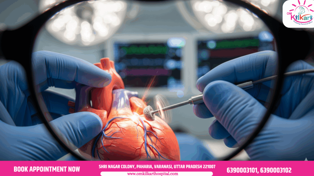
The procedure gets its name from the device used: the Rotablator™. This sophisticated system consists of a console (the control unit) and a catheter-based tool tipped with a tiny, olive-shaped burr. This burr is no ordinary drill bit; it is coated in microscopic diamond crystals, making it one of the hardest substances on earth, perfect for grinding through calcium.
The burr is spun at extremely high speeds—between 140,000 to 200,000 revolutions per minute (RPM)—by a compressed air turbine. It is advanced over a specially designed guidewire (the “RotaWire”) that acts like a track, ensuring the burr stays centered and only grinds the plaque, not the healthy artery wall.
As the diamond-tipped burr rotates at these incredible speeds, it pulverizes the calcified plaque into millions of microscopic particles (smaller than red blood cells). These particles are then safely carried away by the bloodstream and ultimately filtered out by the liver, spleen, and kidneys.
It’s crucial to understand that Rotablation is not a standalone treatment. Its primary purpose is to modify or prepare the artery. By breaking up the concrete-like calcium, it creates a smooth channel, allowing the subsequent balloon to inflate fully and the stent to be deployed optimally and symmetrically.
In essence: Rotablation paves the way for a successful stent placement where standard techniques might fail.
Part 2: The Rotablation Procedure: A Step-by-Step Walkthrough
Understanding the process can alleviate much of the anxiety surrounding a cardiac procedure. Here’s what typically happens from start to finish.
Step 1: Pre-Procedure Preparation
The process begins like any other cardiac catheterization. You will be given a mild sedative to help you relax, but you will remain awake. The area of access—usually the wrist (radial artery) or the groin (femoral artery)—is cleaned and numbed with local anesthetic.
Step 2: Gaining Access and Diagnostic Angiogram
The cardiologist inserts a small sheath into the artery. Through this sheath, a catheter (a thin, flexible tube) is threaded all the way up to the heart’s arteries under X-ray guidance (fluoroscopy). A contrast dye is injected to make the arteries visible on the X-ray screen, providing a clear map of the blockages and their calcification.
Step 3: Delivering the RotaWire
Once the problematic lesion is identified, the cardiologist will carefully navigate the specialized, stainless-steel RotaWire across the calcified blockage. This wire is the foundation for the entire procedure; the burr will travel over it.
Step 4: The “Rota” Run – Advancing the Burr
The diamond-tipped burr is selected based on the size of the artery (usually 1.25mm to 2.5mm in diameter). It is attached to its drive shaft and advanced over the RotaWire until it reaches the beginning of the lesion.
The console is activated, and the burr begins to spin. The cardiologist gently and expertly “pecks” or “dabs” the burr against the calcified segment, advancing it slowly for short periods (typically 15-30 seconds at a time). This intermittent technique prevents overheating and ensures adequate blood flow between runs.
You might hear a faint, high-pitched “whirring” sound from the drive shaft during these runs.
Step 5: Post-Rotablation Angioplasty and Stenting
After the Rotablator has successfully modified the plaque and created a smoother channel, it is removed. The job is only half done. Now, a standard balloon catheter is advanced and inflated to further widen the artery. Finally, a drug-eluting stent (a mesh scaffold that releases medication to prevent re-blockage) is placed in the newly opened section of the artery and expanded.
Step 6: Completion and Removal
A final angiogram is performed to confirm the artery is widely open, blood flow is restored (known as TIMI 3 flow), and the stent is well-apposed to the vessel wall. All catheters and the sheath are removed, and pressure is applied to the access site to prevent bleeding.
The entire procedure can take anywhere from one to three hours, depending on the complexity of the blockages.
Part 3: The Critical Benefits and Advantages of Rotablation
Why go through this more complex procedure? The benefits in complex cases are significant and often make the difference between success and failure.
- Enables Successful Stent Deployment: This is its primary benefit. In heavily calcified lesions, a stent may not fully expand. An underexpanded stent is a major risk factor for stent thrombosis (a deadly blood clot) and in-stent restenosis (re-narrowing). Rotablation virtually eliminates this risk by ensuring the stent can be expanded to its intended diameter.
- Improves Safety and Efficacy of Balloon Angioplasty: Trying to force a high-pressure balloon through a rigid, calcified lesion can lead to vessel dissection (a tear in the artery wall) or even vessel rupture. By pre-treating the lesion, Rotablation allows for balloon inflation at lower pressures, drastically reducing this risk.
- Facilitates Complex Procedures: It is often the only way to perform certain interventions, such as:
- Treating Bifurcation Lesions: Blockages at a branch point in an artery.
- Facilitating Lithotripsy: Sometimes, Rotablation is used to create a small channel to allow a Shockwave Lithotripsy balloon to be delivered to the lesion.
- Expanding Underexpanded Stents: It can be used to grind the calcium behind a stent that failed to expand during a previous procedure.
- “Debulking” the Plaque: While not its main goal, the mechanical removal of plaque volume can lead to a larger final lumen (channel) size after stenting, which is associated with better long-term patency.
- A Proven, Time-Tested Technology: With decades of clinical use and data, Rotablation has a well-established safety profile in the hands of experienced operators.
Part 4: Who is a Candidate? Indications and Treatments
Rotablation is a highly specialized tool, not for every blockage. Patient selection is paramount.
The Primary Indication: Severe Coronary Artery Calcification
The ideal candidate is a patient whose angiogram or intravascular imaging (like IVUS or OCT) reveals a lesion that is:
- Un-dilatable: A balloon catheter cannot be inflated across it even with high pressure.
- Un-crossable: A balloon or stent cannot even be delivered across the lesion.
- Likely to cause stent failure: Where the cardiologist predicts poor stent expansion based on the calcium seen on imaging.
Specific Clinical Scenarios:
- In-Stent Restenosis (ISR) with Calcium: When a previously placed stent has re-narrowed due to calcified tissue growth.
- Ostial Lesions: Blockages located at the very origin of an artery (e.g., aorta-ostial lesions), which are often fibrotic and calcified.
- Long, Diffuse Calcified Lesions: Extensive calcium deposits over a long segment of an artery.
- Moderately Calcified Lesions in Tortuous Vessels: To make the path straighter and safer for stent delivery.
Contraindications: When Rotablation is Not Suitable
The procedure is not risk-free and is avoided in certain situations:
- Presence of a Thrombus (Blood Clot): The rotating burr could break the clot loose, causing an embolism.
- Vein Graft Lesions: Saphenous vein grafts used in bypass surgery are often soft and friable; Rotablation could cause significant debris embolization.
- Poor LV Function or Hemodynamic Instability: The procedure can cause transient slow heart rhythm, which an unstable patient may not tolerate.
- Allergy to Contrast Dye or RotaWire Components.
The decision to use Rotablation is made by a heart team after a thorough review of the patient’s anatomy, clinical status, and risks.
Part 5: Potential Risks and Complications
As with any invasive cardiac procedure, Rotablation carries risks. However, in experienced centers, the rate of major complications is low (<2%).
- Bradycardia (Slow Heart Rate): The most common issue. The high-speed burr can stimulate the vagus nerve, causing a transient, often profound, slowing of the heart rate. This is why all patients are pre-treated with atropine and often have a temporary pacemaker placed as a precaution.
- Coronary Artery Spasm: The rotation can cause the artery to constrict. This is managed with medications like nitroglycerin.
- No-Reflow Phenomenon: Microscopic debris can block small downstream blood vessels, preventing blood from re-perfusing the heart muscle tissue. Treated with vasodilators.
- Perforation or Dissection: A rare but serious risk of the burr damaging the artery wall.
- Burr Entrapment: The burr can get stuck if advanced too aggressively. Advanced techniques are required to free it.
- Embolization: While particles are microscopic, they can occasionally cause issues in patients with pre-existing poor kidney function.
The key to mitigating these risks lies in the operator’s experience, careful patient selection, and meticulous technique.
Part 6: The Recovery Timeline: What to Expect After the Procedure
Recovery from a Rotablation procedure is very similar to recovery from a standard angioplasty and stenting.
Immediately After the Procedure (First 4-6 Hours):
You will be moved to a recovery room for close monitoring. The nursing staff will check your vital signs, the access site (wrist or groin) for bleeding, and your heart rhythm. You will need to lie flat, especially if the groin was used, to prevent bleeding. You will be encouraged to drink fluids to help flush the contrast dye from your system.
Hospital Stay (Usually 1-2 Nights):
Most patients stay in the hospital for observation. During this time, your cardiac team will ensure you are stable and adjust your medications. You will be started on or continued on dual antiplatelet therapy (DAPT) – usually Aspirin and a drug like Clopidogrel (Plavix), Ticagrelor (Brilinta), or Prasugrel (Effient). Adherence to DAPT is absolutely critical to prevent blood clots from forming on the newly placed stent.
First Week at Home:
- Activity: Take it easy. Avoid heavy lifting (nothing over 10 pounds), strenuous exercise, and driving for the first few days to a week. Walking is encouraged and beneficial.
- Access Site Care: Keep the area clean and dry. You can usually shower after 24-48 hours. Watch for signs of infection: redness, swelling, drainage, or fever.
- Medications: Take all prescribed medications exactly as directed. Do not stop your blood thinners unless explicitly told to do so by your cardiologist.
Long-Term Recovery (1-6 Months and Beyond):
- Cardiac Rehabilitation: This is a strongly recommended, supervised program of exercise training, education, and counseling. It is proven to reduce mortality, improve recovery, and help you return to an active life.
- Lifestyle Modifications: The procedure fixes a blockage, but it does not cure the underlying heart disease. Long-term success depends on you:
- Heart-Healthy Diet: Mediterranean or DASH diet, low in saturated fats, salt, and processed sugars.
- Regular Exercise: Aim for at least 150 minutes of moderate exercise per week.
- Smoking Cessation: This is non-negotiable.
- Manage Other Conditions: Strict control of blood pressure, cholesterol, and diabetes.
- Follow-Up Appointments: You will have regular check-ups with your cardiologist to monitor your progress, manage medications, and ensure your heart remains healthy.
Part 7: The Future of Plaque Modification
Rotablation remains a cornerstone for treating severe calcium, but it’s not the only technology. A newer technique called Intravascular Lithotripsy (IVL), commercially known as the Shockwave Medical system, has emerged.
This technology uses sonic pressure waves (similar to those used to break up kidney stones) to crack calcium from within an angioplasty balloon, without the high-speed rotation. Studies show it is highly effective and has an excellent safety profile.
The choice between Rotablation, Lithotripsy, or other techniques like orbital atherectomy depends on the specific characteristics of the calcium, the operator’s expertise, and institutional availability. Often, these tools are complementary, and interventional cardiologists are skilled in using the right tool for the right job.
Conclusion: A Precision Tool for a Tough Problem
Rotational Atherectomy, or Rotablation, is a testament to medical innovation. It is a highly specialized, life-enabling procedure that allows interventional cardiologists to successfully treat some of the most challenging coronary blockages. By using a diamond-tipped burr spinning at incredible speeds, it masterfully applies the principle of differential cutting to ablate rock-hard calcium while preserving healthy tissue.
While it carries specific risks, its benefits in facilitating optimal stent placement in complex cases are undeniable. For patients facing the prospect of open-heart surgery due to severe calcification, Rotablation can often provide a less invasive, highly effective alternative.
If you or a loved one is diagnosed with severely calcified coronary artery disease, have a detailed discussion with your cardiologist and interventionalist. Ask about the nature of your blockage, the treatment options available (including Rotablation and Lithotripsy), and the experience of your medical team. An informed patient is an empowered partner in their own heart health journey.